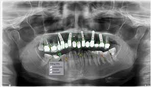


Orthopantomogram (OPG), or panoramic dental X-ray, is a transformative tool in modern dentistry, offering a comprehensive view of the oral cavity in a single image. Hamintour, a trusted name in medical tourism, integrates this advanced diagnostic technology to ensure precise dental care for international patients visiting Iran. This guide explores the technical brilliance, clinical applications, and future potential of OPG, enriched with the latest insights and practical examples.
What is a Dental OPG?
An OPG is a panoramic radiograph capturing the entire oral structure—teeth, upper and lower jaws (maxilla and mandible), temporomandibular joints (TMJ), and surrounding tissues—in a single, two-dimensional image. Unlike intraoral X-rays, which focus on specific teeth, OPG provides a holistic view, making it indispensable for diagnosing complex dental conditions.
Key Features of OPG
-
Comprehensive Imaging: Visualizes all teeth and jaw structures in one scan.
-
Non-Invasive: Quick and comfortable for patients.
Table of Contents -
Low Radiation: Delivers 5.5–22 μSv, equivalent to 1–3 days of natural background radiation (PubMed, 2023).
-
Cost-Effective: Reduces the need for multiple X-rays, saving time and money.
Historical Evolution of OPG
The concept of panoramic imaging dates back to 1933, when Dr. Hisatugu Numata patented an early design. The breakthrough came in the 1950s with Dr. Yrjö Paatero’s Orthopantomograph, the first commercially viable panoramic X-ray machine. Today, digital advancements and AI integration have elevated OPG’s precision and accessibility, making it a cornerstone of dental diagnostics globally, including at Hamintour’s partner clinics in Iran.
How OPG Technology Works
OPG machines use synchronized movements to capture detailed images:
-
X-Ray Tube and Receptor: The tube emits a collimated X-ray beam while the receptor rotates around the patient’s head.
-
Focal Trough: Structures within this plane are sharply imaged, while others are blurred.
-
Digital Processing: Advanced sensors and software produce high-resolution images in seconds.
This process, completed in 12–20 seconds, ensures minimal discomfort and rapid results, a standard upheld by Hamintour’s partner facilities in Iran.
Clinical Applications of OPG
OPG’s versatility makes it a vital tool across various dental specialties. Below are its primary applications, with real-world examples:
General Dentistry
-
Tooth Development: Identifies eruption patterns, aiding pediatric dentistry.
-
Impacted Teeth: Detects wisdom teeth misalignment, common in 30% of adults (WHO, 2024).
-
Periodontal Health: Assesses bone loss, critical for diagnosing periodontitis, affecting 19% of the global population (Journal of Periodontology, 2023).
Case Example: A 28-year-old patient visited a Hamintour-affiliated clinic in Tehran for jaw pain. An OPG revealed an impacted lower wisdom tooth, leading to a planned extraction and follow-up care, ensuring quick recovery.
Orthodontic Planning
-
Tooth Positioning: Evaluates alignment for braces or aligners.
-
Jaw Relationships: Assesses skeletal discrepancies for treatment planning.
Case Example: A teenager in Shiraz, guided by Hamintour, underwent an OPG scan revealing crowding in the upper jaw. Orthodontic treatment was planned, reducing treatment time by 20% due to precise diagnostics.
Surgical and Implant Planning
-
Wisdom Tooth Extraction: Identifies nerve proximity to minimize risks.
-
Dental Implants: Assesses bone density, crucial for 95% implant success rates (International Journal of Oral Implantology, 2024).
Case Example: A 45-year-old patient sought dental implants through Hamintour. OPG imaging confirmed sufficient bone volume in the mandibular region, enabling successful implant placement and a zirconia crown restoration.
Pathological Detection
-
Cysts and Tumors: Identifies abnormalities like ameloblastomas, which occur in 1% of oral tumors (Oral Oncology, 2023).
-
TMJ Disorders: Detects joint issues, affecting 5–12% of adults (American Dental Association, 2024).
Case Example: An OPG at a Hamintour partner hospital identified a mandibular cyst in a 35-year-old patient, leading to early surgical intervention and full recovery.
Advantages of OPG in Dental Care
-
Holistic View: Captures the entire oral cavity, reducing the need for multiple scans.
-
Patient-Friendly: Non-invasive, with scans taking less than 20 seconds.
-
Radiation Safety: Lower dose than full-mouth X-ray series, aligning with ALARA principles (As Low As Reasonably Achievable).
-
Efficiency: Immediate digital results enhance treatment planning.
Hamintour ensures patients access state-of-the-art OPG technology at accredited clinics, combining affordability with quality care.
Limitations of OPG
Despite its benefits, OPG has constraints:
-
2D Imaging: May miss fine details, requiring supplementary 3D scans like CBCT.
-
Distortion Risk: Incorrect positioning can affect image accuracy.
-
Not for Small Cavities: Intraoral X-rays are better for detecting early caries.
Clinical Tip: Hamintour’s partner dentists recommend combining OPG with targeted intraoral X-rays for comprehensive diagnostics, ensuring no detail is overlooked.
The OPG Procedure: What to Expect
Preparation
-
Remove Metal Objects: Jewelry, glasses, or dentures can interfere with imaging.
-
Positioning: Patients bite on a plastic block while alignment lights ensure accuracy.
-
Radiation Protection: Lead aprons and thyroid collars are standard, minimizing exposure.
During the Scan
-
The machine rotates around the head for 12–20 seconds.
-
Patients remain still to avoid image distortion.
-
Digital sensors capture and process the image instantly.
Hamintour’s coordinators guide patients through this process, ensuring comfort and clarity at every step.
Radiation Safety in OPG
OPG’s radiation dose is minimal, ranging from 5.5–22 μSv, compared to 35–150 μSv for a full-mouth series (Radiology, 2023). Safety measures include:
-
Patient Protection: Lead aprons and thyroid collars.
-
Operator Safety: Shielded barriers and regular equipment calibration.
-
Digital Advantage: Reduces radiation by up to 30% compared to analog systems (Journal of Dental Research, 2024).
Hamintour prioritizes clinics adhering to strict radiation safety protocols, ensuring patient and staff well-being.
Advances in Digital OPG Technology
Digital Sensors
-
Enhanced Clarity: Higher resolution than traditional film X-rays.
-
Instant Results: Images are available within seconds for immediate analysis.
-
Eco-Friendly: Eliminates chemical processing.
AI Integration
-
Automated Analysis: AI algorithms detect caries and cysts with 90% accuracy (Journal of Dental Imaging, 2024).
-
Treatment Planning: Suggests optimal implant placement or orthodontic adjustments.
Example: A Hamintour patient in Mashhad benefited from AI-enhanced OPG analysis, which identified early periodontal bone loss, enabling preventive treatment.
Future of OPG in Dentistry
Emerging trends promise to elevate OPG’s role:
-
3D Hybrid Imaging: Combines OPG with CBCT for detailed diagnostics.
-
AI Diagnostics: Machine learning improves pathology detection, reducing diagnostic errors by 15% (Nature Dentistry, 2025).
-
Tele-Dentistry: Digital OPGs enable remote consultations, a growing trend in Iran’s medical tourism sector.
Hamintour stays ahead by partnering with clinics adopting these innovations, ensuring patients receive cutting-edge care.
Why Choose Hamintour for Dental Care in Iran?
Hamintour transforms dental tourism by offering:
-
Affordable Excellence: Dental procedures in Iran cost 50–70% less than in Western countries (e.g., implants at $800 vs. $3,000 in the US).
-
Comprehensive Packages: Include OPG diagnostics, surgery, hotel, visa, and interpreter services.
-
Trusted Clinics: Partnered with JCI-accredited facilities in Tehran, Shiraz, and Mashhad.
Patient Story: Maria, a 40-year-old from the UK, chose Hamintour for dental implants. Her OPG scan revealed optimal bone structure, and she received two implants and a crown for $2,500, saving 60% compared to UK prices.
FAQs About Dental OPG
What is the difference between OPG and intraoral X-rays?
OPG provides a panoramic view of the entire oral cavity, while intraoral X-rays focus on specific teeth for detailed imaging.
Is OPG safe for pregnant women?
OPG is generally avoided during pregnancy unless essential, with lead aprons used for protection (American College of Radiology, 2024).
How often should I get an OPG?
OPG is recommended based on clinical need, not routine screening, typically every 2–5 years for adults (ADA, 2024).
Can OPG detect all dental issues?
OPG excels in broad diagnostics but may require supplementary imaging for small cavities or soft tissue issues.
Why choose Hamintour for OPG diagnostics in Iran?
Hamintour partners with top-tier clinics, offering affordable, high-quality OPG imaging with seamless travel and treatment coordination.